Anion Gap Calculator for Physicians: Clinical Interpretation Guide
What the Anion Gap Measures
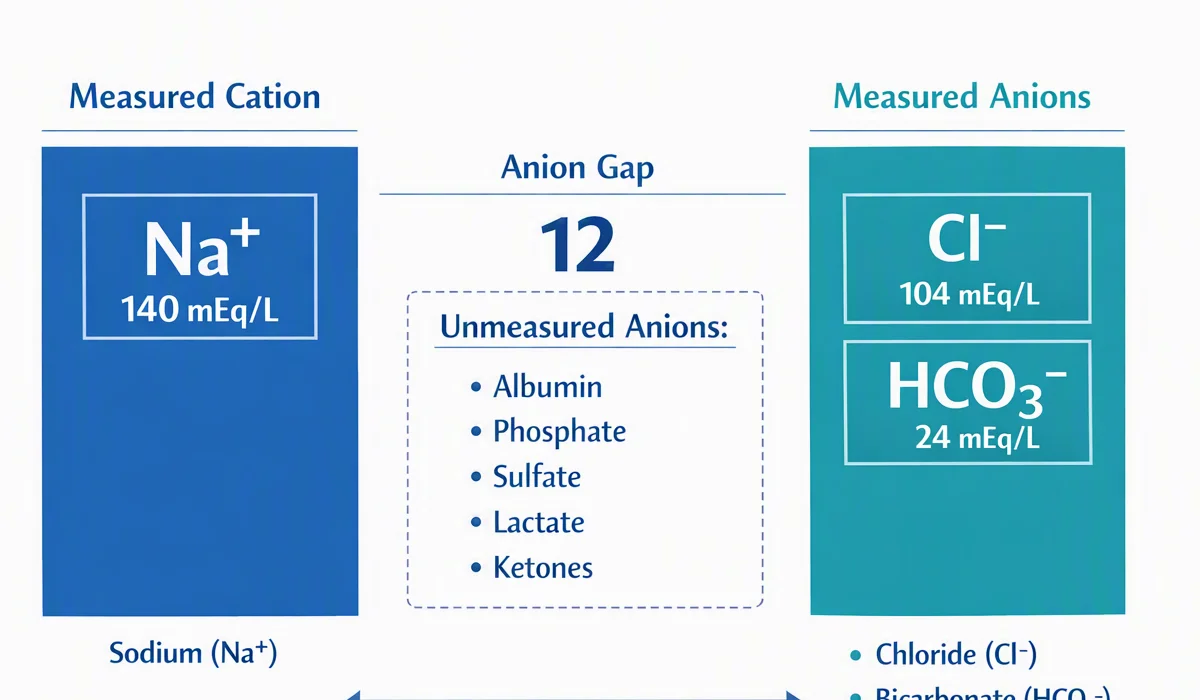
The anion gap is a valuable lab-based estimate used in point-of-care and formal laboratory interpretation to help reveal hidden shifts in serum electrolytes. It reflects the balance between routinely measured cations and anions, most commonly serum sodium, serum chloride, and serum bicarbonate, with serum potassium sometimes included depending on the formula used. Clinically, it functions as a diagnostic marker for acid-base disorder analysis, especially when metabolic acidosis is suspected.
The main value of the anion gap is that it estimates the burden of unmeasured anions in the blood chemistry. These may include lactate, ketones, sulfate, phosphate, and various toxins. A rise in the gap usually indicates a metabolic derangement that has introduced acid equivalents or reduced bicarbonate without a proportional increase in chloride. This is why the anion gap remains central to acid-base balance assessment and clinical assessment in acutely ill patients.
From a pathophysiology standpoint, the calculation is grounded in electroneutrality. Because standard chemistry panels do not measure every ion, the gap helps infer hidden contributors to systemic acidosis. In practice, the result can suggest whether the body is experiencing a chloride-driven process, a bicarbonate deficit, or accumulation of acids from toxic ingestion, hypoperfusion, or impaired renal excretion.
Most clinicians use the anion gap as part of a broader differential diagnosis. It is not a standalone diagnosis, but a structured way to interpret the pattern of electrolyte panel results in context. When combined with pH, bicarbonate, and the rest of the serum electrolytes, it can rapidly narrow the likely cause of acidosis and guide the next diagnostic step.
How to Operate an Anion Gap Calculator
A Anion Gap Calculator streamlines the anion gap formula and reduces arithmetic errors during urgent clinical assessment. Most calculators ask for serum sodium, serum chloride, and serum bicarbonate; a few also request serum potassium. The calculator then returns the calculated gap and often indicates whether the result matches the expected reference range.
Without potassium, the formula is:
Anion gap = sodium − (chloride + bicarbonate)
If potassium is included, the equation becomes:
Anion gap = sodium + potassium − (chloride + bicarbonate)
To use it well, enter the exact lab values from the same sample whenever possible. Combining results from different draws can distort the result, especially in rapidly changing clinical states. The calculator is most useful when the chemistry panel, acid-base data, and bedside findings are interpreted together rather than in isolation.
During routine care, the calculator should be paired with the broader picture of blood chemistry. For instance, a low bicarbonate with a high anion gap supports high-gap acidosis, but the source of the acid still requires further workup. If the gap is normal despite acidosis, the calculator may point toward a chloride-rich process or another type of acid-base disorder.
These practical tips improve accuracy:

- Use the same reporting units consistently.
- Verify whether potassium is part of the local formula.
- Check for unexpected lab artifacts before overinterpreting a borderline value.
- Assess the albumin level if the result seems discordant with the clinical picture.
The most useful Anion Gap Calculator output is not the number alone, but the interpretation pathway it triggers. A calculated gap that is unexpectedly high should prompt a search for lactic acidosis, ketoacidosis, renal failure, or toxic alcohols. A normal gap with persistent acidemia raises concern for gastrointestinal or renal bicarbonate loss, among other etiologies.
Normal vs Elevated Anion Gap
The normal anion gap is best understood as a span rather than a single fixed number because it changes by testing method, analyzer platform, and whether potassium is included. This is why the reported reference range from the local lab should guide interpretation. A value in the anticipated range does not automatically exclude disease; it simply implies that the current acid-base pattern does not show a substantial burden of unmeasured ions.
An elevated gap is often the clue that redirects the differential toward high anion gap metabolic acidosis. In this setting, the accumulation of unmeasured acids lowers bicarbonate as chloride does not rise enough to fully offset the charge change. The resulting pattern is a hallmark of a clinically important acid-base disorder and often warrants urgent evaluation for reversible causes.
By contrast, a normal anion gap in the presence of low bicarbonate typically points toward a hyperchloremic process. Here, chloride rises as bicarbonate falls, preserving electroneutrality and keeping the gap relatively stable. This pattern is frequently seen in diarrhea or renal tubular defects, but it can also be altered by saline administration and other treatment-related shifts.
Understanding whether a patient has metabolic acidosis requires more than a single chemistry result. A low bicarbonate with normal pH may reflect compensation, mixed disorders, or timing issues. Likewise, the anion gap may be elevated before pH becomes severely abnormal if respiratory compensation is intact. This is why interpretation should always include the full acid-base picture, not just the numeric gap.
Revised Anion Gap and Albumin Correction
Hypoalbuminemia is a major reason a measured gap can look falsely reassuring or only mildly elevated. Because albumin is a principal unmeasured anion, reduced albumin reduces the baseline gap and may hide clinically meaningful acid accumulation. This is the reason for the corrected anion gap and broader anion gap correction strategy.
Adjusted albumin interpretation helps avoid missing a high-gap process in critically ill patients. When albumin is low, the measured gap should be interpreted together with a correction factor rather than as a raw number alone. The goal is not to “normalize” the lab value cosmetically, but to uncover the true magnitude of the underlying electrolyte imbalance.
Many clinicians use an albumin-adjusted approach when the clinical suspicion for acidosis is high but the initial gap is mild. This matters in sepsis, liver disease, malnutrition, nephrotic states, and other conditions associated with hypoalbuminemia. Without correction, a dangerous acid load may be missed.
A practical approach is to check the albumin whenever the chemistry profile does not align with the bedside picture. If the albumin is low, recalculating the corrected anion gap can reclassify a borderline result into a clearly abnormal one. This boosts diagnostic sensitivity for hidden metabolic acidosis and helps align the lab interpretation with the patient’s physiology.
Albumin correction is especially important when the differential includes toxic or ischemic etiologies. In those situations, a falsely reassuring value can delay treatment. Using correction routinely in the appropriate context decreases the chance of missing a significant metabolic derangement.
Frequent Causes of elevated anion gap metabolic acidosis
High anion gap metabolic acidosis indicates the accumulation of acid precursors or acid https://anion-gap-results619.cavandoragh.org/anion-gap-calculator-for-kidney-disease-evaluation precursors that create unmeasured ions. The leading causes in clinical practice include lactic acidosis, ketoacidosis, renal failure, and toxic alcohols. Each has separate mechanisms, but they produce the same laboratory pattern: a drop in bicarbonate with a elevated gap.
Lactic acidosis arises when tissue hypoperfusion, low oxygen levels, mitochondrial dysfunction, or severe metabolic stress drives lactate production faster than elimination. It is common in shock states, sepsis, seizures, and marked anemia. The anion gap may rise quickly, making it an initial clue of occult hypoperfusion and worsening systemic acidosis.
Ketoacidosis occurs when ketone production outpaces utilization and elimination. Diabetic ketoacidosis is the best-known form, but starvation and alcohol-related states can also produce a significant gap. The degree of acidemia varies, and the final pattern depends on insulin status, volume depletion, and compensatory respiratory response.
Renal failure contributes by reducing acid excretion and allowing retention of sulfate, phosphate, and other acidic solutes. In advanced kidney dysfunction, the gap may rise progressively or considerably depending on the degree of retained acids and concurrent therapies. A worsening gap in renal failure should prompt review of the trajectory and any superimposed lactic or ketoacidotic process.
Toxic alcohols remain a critical diagnosis not to miss. Ethylene glycol, methanol, and related substances can cause marked high-gap acidosis through metabolite accumulation. The anion gap may be elevated before the osmolar gap closes, so repeat testing and detailed history are essential. In any suspected toxic ingestion, the gap is a valuable clue but never definitive on its own.
Additional causes can include salicylates, rhabdomyolysis, and certain inborn errors, but the major practical framework remains the same: identify the acid source, gauge severity, and determine whether urgent antidotal or supportive treatment is needed. The calculator is best viewed as an entry point into a structured diagnostic differential.
Typical Causes of Normal Gap Metabolic Acidosis
A normal gap metabolic acidosis is usually a chloride-mediated process in which bicarbonate is lost or substituted in a way that maintains the measured gap. The main two clinical causes are diarrhea and tubular kidney acidosis. In these cases, the lab pattern often points toward chloride-associated acidosis.
Diarrhea causes direct loss of bicarbonate from the gastrointestinal tract. As bicarbonate falls, chloride rises to maintain electroneutrality, resulting in a relatively normal gap. The history is often key, but the chemistry panel can confirm the physiology when symptoms are incomplete or communication is limited.
Renal tubular acidosis reflects impaired renal acid handling or bicarbonate reclamation. Different subtypes affect distal acid secretion or proximal bicarbonate reabsorption, but the final blood chemistry can be similar: low bicarbonate, relatively preserved anion gap, and often hyperchloremia. Recognition matters because management may differ by subtype and chronicity.
Chloride-associated acidosis may also appear after substantial chloride-rich fluid administration or in settings where bicarbonate is replaced by chloride at the kidney or gut level. This is an important reminder that the gap is a pattern recognition tool, not a diagnosis in itself. Correlation with iatrogenic exposures, kidney function, and GI losses is essential.
In all normal-gap states, the question is not simply “why is the gap normal?” but “what mechanism is replacing bicarbonate with chloride or preventing acid excretion?” That physiologic question guides the next step in workup and management.
Clinical Analysis and Further Steps
Assessing the anion gap in practice starts with the acid-base data and then broadens into the broader clinical context. An arterial blood gas is often the best next study when the chemistry panel suggests important abnormality. The blood gas helps establish the pH, evaluate respiratory compensation, and identify mixed disorders that are not obvious from chemistry alone.
The delta gap is important when the measured gap is elevated. It compares the rise in the anion gap with the fall in bicarbonate to determine whether a second metabolic process is present. A disproportionately large gap rise relative to bicarbonate decline may suggest concurrent metabolic alkalosis; a smaller-than-expected change may indicate a mixed high-gap and normal-gap acidosis. This step sharpens diagnosis beyond the initial screening value.
Base status also matters. A significant base deficit supports clinically relevant acid burden and helps track severity over time. In shock, sepsis, or toxin exposure, it may provide a useful serial indicator alongside serial lactate, renal function, and repeated electrolyte measurements.
A practical workflow is:
- Check the chemistry values and calculate the gap.
- Assess albumin and apply correction if needed.
- Obtain the arterial blood gas for pH and compensation.
- Use the delta gap when the gap is elevated.
- Look for the underlying cause based on the pattern and clinical context.
In a patient with high-gap acidosis, prioritize rapid identification of lactic acidosis, ketoacidosis, renal failure, and toxic alcohols. In a normal-gap acidosis, turn to diarrhea, renal tubular acidosis, and other sources of bicarbonate loss. If the picture is mixed or uncertain, repeat the labs, review prior values, and re-evaluate the trajectory rather than anchoring to one result.
Constraints of Anion Gap Calculation
Even with its usefulness, the anion gap has notable drawbacks. Laboratory variation can alter the expected reference range between institutions and analyzers, so a number that is “normal” in one setting may be somewhat abnormal in another. This is one reason local reference intervals should always be used for interpretation.
Testing error is another challenge. Small inaccuracies in sodium, chloride, or bicarbonate can materially change the calculated result, especially when the gap is borderline. Preanalytic issues, sample handling, and analyzer-specific bias all affect the output. If the value does not fit the clinical picture, repeat testing is often appropriate before making major decisions.
Another major limitation is the influence of unmeasured cations. Lithium, marked hypermagnesemia, and other cation excess states can lower the gap and obscure underlying acidosis. Likewise, shifts in calcium or paraproteins may alter interpretation in select cases. The calculator cannot fully account for these possibilities without broader clinical data.
Most importantly, the anion gap must always be interpreted in clinical context. The same value can mean different things depending on kidney function, albumin, ventilation, perfusion, medications, and timing. The calculator supports clinical evaluation, but it does not replace judgment, trend review, or diagnosis by cause.
If used correctly, the tool is a powerful aid to interpretation of laboratory results. If taken on its own, it can mislead. The best practice is to integrate the gap with symptoms, serial chemistry, gas exchange, and the broader physiologic picture.
Frequently Asked Questions
How is the anion gap calculated?
The most commonly used anion gap equation is sodium minus the sum of chloride and bicarbonate. Some providers include potassium as well, depending on local convention and the calculator used. The key is to apply the same method consistently and interpret the result against the relevant laboratory reference range.
What is the reason for albumin correction important while interpreting the anion gap?
Albumin serves as a major nonmeasured anion, so hypoalbuminemia reduces the apparent gap and can mask a true acid burden. A corrected anion gap or albumin-adjusted interpretation boosts detection of hidden high-gap states and decreases the risk of underestimating metabolic acidosis.
What does a high anion gap indicate from a clinical standpoint?
A high gap usually reflects accumulation of unmeasured anions from conditions such as lactic acidosis, ketoacidosis, renal failure, or toxic alcohols. It is a pointer to high anion gap metabolic acidosis and should prompt a focused search for the underlying cause.
Is it possible for the anion gap be normal in metabolic acidosis?
Yes. A normal anion gap can occur in metabolic acidosis when bicarbonate is lost and replaced by chloride, producing hyperchloremic acidosis. Common causes include diarrhea and renal tubular acidosis, both of which can lower bicarbonate without widening the gap.
What are the main limitations of using an anion gap calculator?
The main limitations include lab variation, measurement error, the influence of unmeasured cations, and dependence on clinical context. The calculator is useful for screening and pattern recognition, but it cannot replace a full acid-base evaluation, including the arterial blood gas, albumin review, and cause-specific differential diagnosis.